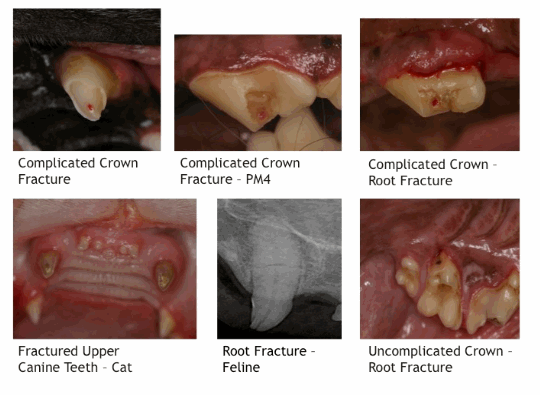
Fractured Pet Teeth
Fractured teeth are common in dogs and cats and usually caused by either trauma to the head or from pets chewing on inappropriately hard objects such as bones. Often fractured teeth go unnoticed by the owners unless they directly observe the injury when it takes place. Veterinarians and technicians often identify fractured teeth incidentally while performing routine oral examinations.
Physical and radiographic evaluations are essential to determine the best treatment option for an individual fractured tooth. Conscious oral exams performed in the exam room are of limited value. A complete, more thorough evaluation must be performed while the patient is anesthetized.
Once the patient is anesthetized, physical evaluation can be performed and dental radiographs obtained. A pointed dental explorer is used to probe the dental tissues for loose fragments, cracks, and to assess whether the fracture has exposed the pulp chamber (complicated fracture). A periodontal probe is used to evaluate the extent to which a slab fracture extends below the gingival margin. Transillumination can help reveal vertical fractures as well as determine tooth vitality. A vital tooth will have a translucent appearance while a non-vital tooth may appear opaque. Dental radiographs are necessary to complete any tooth evaluations, and to directly assess whether or not there are root fractures or if an apical periodontitis is present.
Enamel Fractures: A simple crown fracture involving just the enamel may only require smoothing the enamel with a fine diamond bur in a water-cooled, high-speed hand piece.
Uncomplicated Crown Fractures: These are defined as fractures that include both enamel and dentin layers of the tooth, however, there is no pulp exposure. Treatment goals are to protect and restore the tooth by using layers of bonded dental sealants and composites. After the tooth is smoothed and the enamel beveled, the fractured tooth is cleaned, polished with flour of pumice, etched, and treated with a bonded dental sealant. A composite filling material can then be placed over the fracture to restore the tooth and provide an additional protective layer. Some uncomplicated fractures may actually occur at a depth where a “near pulp exposure” has occurred (pink spot in the dentin over the area of the pulp chamber). For treatment, an additional protective layer for the pulp may be indicated. Once the near pulp exposure is treated, the fractured tooth can be restored as previously described. Even uncomplicated crown fractures may traumatize the pulp to the degree that it eventually becomes non-vital (necrotic). Follow-up radiographs are indicated.
Complicated Crown Fractures: These are defined as fractures that extend into and expose the pulp. If the pulp exposure is recent (24-48 hours since exposure in a mature dog, or up to 2 weeks exposure in a dog less than 18 months of age), vital pulp therapy (VPT, partial pulpectomy, and directly medicating the pulp) may be a treatment option. After performing VPT, the tooth is restored with adhesives and composites as described above. If the fractured tooth with pulp exposure does not meet the criteria for partial pulpectomy and pulp capping, then root canal therapy is indicated before restoring the fractured tooth. Any non-vital tooth will become infected. It is only a matter of time. Because of this, from the patient’s perspective, the “wait and see” treatment option approach is incorrect and inappropriate. The pulp must be removed and is done so by either extracting the tooth or having a root canal procedure performed.
Crown-Root Fractures: This type of fracture is often encountered with the classical “slab-fracture” of the upper 4th premolar in dogs. In these cases, the root fracture component disrupts the normal gingival/periodontal attachment around the tooth. This predisposes to focal, chronic periodontitis problems. Therefore, treatment decisions for the tooth need to reflect consideration for the ongoing periodontal health of the tooth.

