
Tooth Resorption
Tooth resorption is a relatively common and painful condition in cats. It can occur in dogs as well, although it is not as common. These lesions used to be referred to as “neck lesions” due to their frequent location at the “neck” of the tooth, where the gingiva meets the crown. They have also been called FORLs (feline odontoclastic resorptive lesions) or feline caries, but this is a misnomer because there have been no reports of cavities in cats. In tooth resorption, odontoclasts, cells that are normally present around the root of a tooth, get activated (for unknown reasons) and begin “eating away” at the tooth substance. This process will continue and eventually affect all parts of the tooth including the crown, dentin, and root canal. This causes inflammation and pain when the lesion is exposed to the oral cavity.
What causes tooth resorption?
We do not know what causes tooth resorption at this time and to date, no studies have been able to elucidate the etiology of this disease in cats. We have seen evidence of tooth resorption in skulls dating back to the 13th century, so it is certainly not a new disease, but it appears that there has been a sharp increase in prevalence over time.
Diagnosis of tooth resorption?
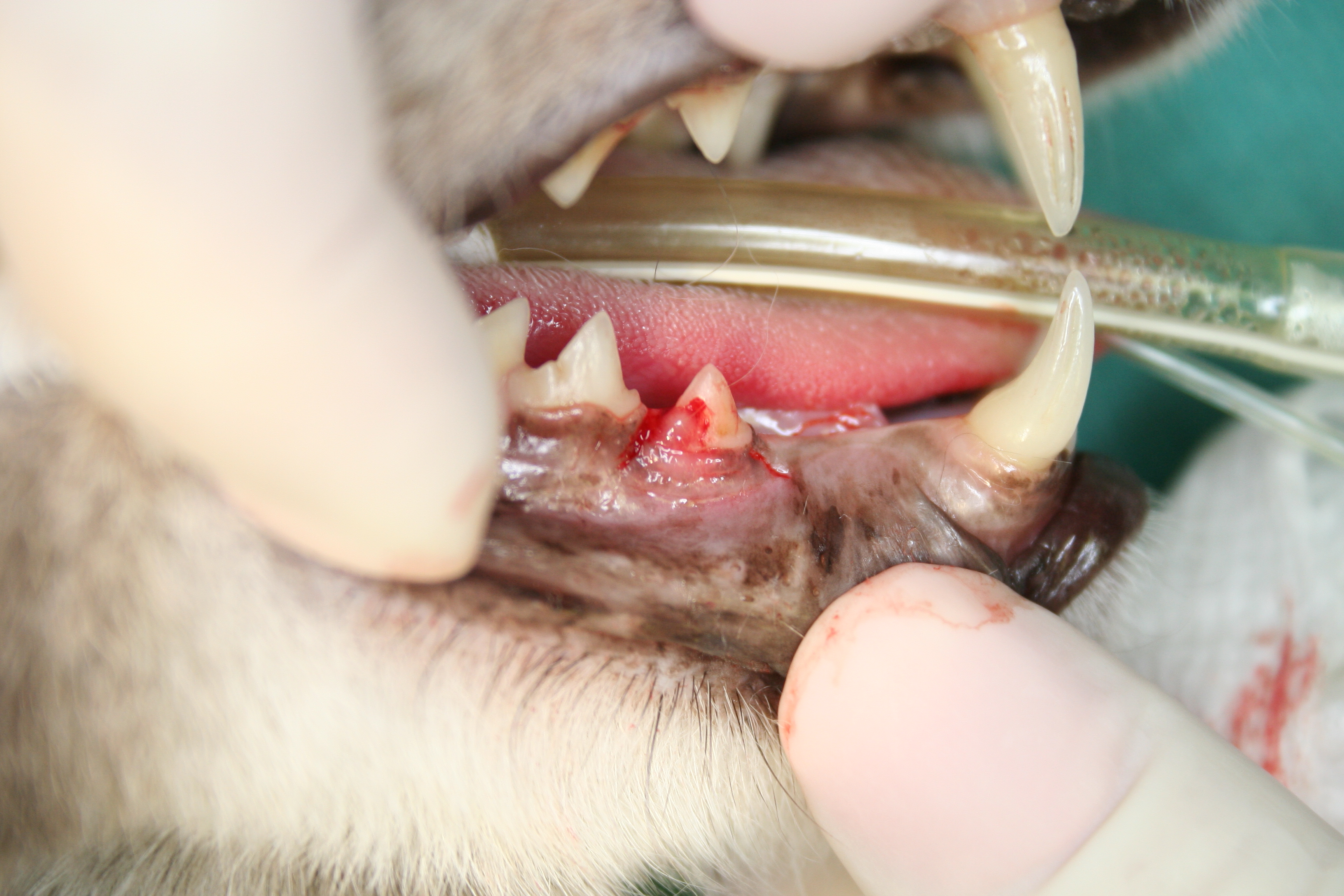
Cats are extremely tolerant of pain and will rarely show obvious signs of oral discomfort unless it is extreme. Because of this, tooth resorption is most commonly diagnosed incidentally on general examination. The mandibular third premolar is the most commonly affected tooth and should be monitored closely on awake examination when possible. If this tooth is missing or affected by resorption, full-mouth x-rays and oral examination should be performed as it is likely that other teeth will be affected as well. Other signs of resorption on awake examination include a visible hole in the tooth and/or inflamed gingiva “growing up” over the surface of the crown.
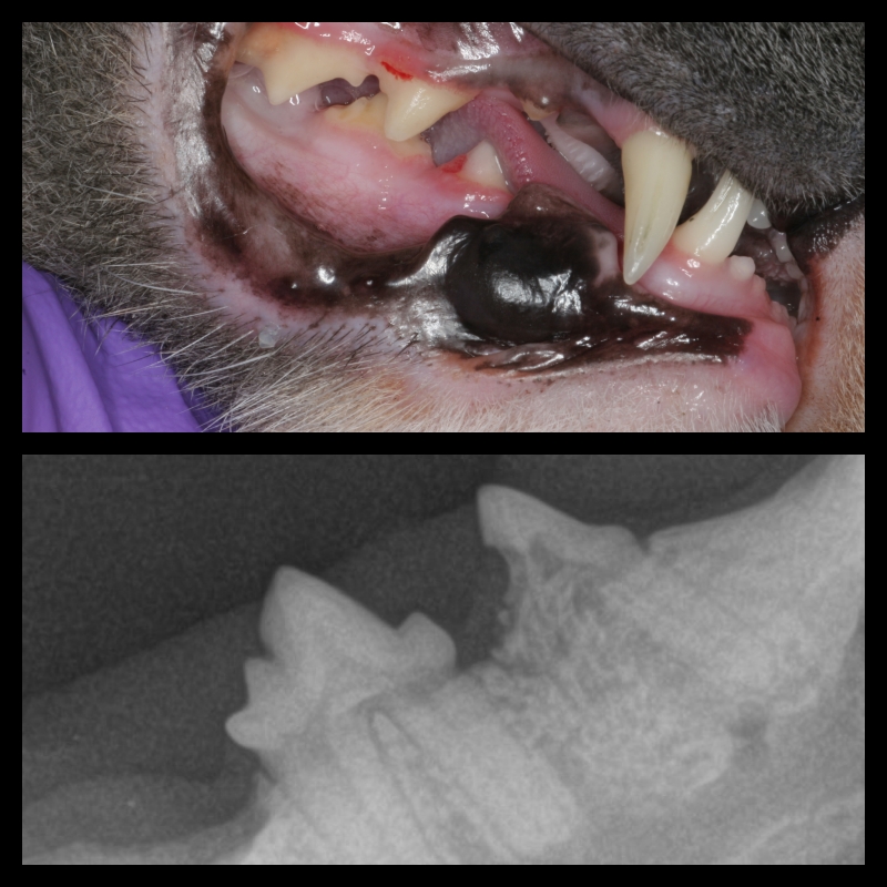
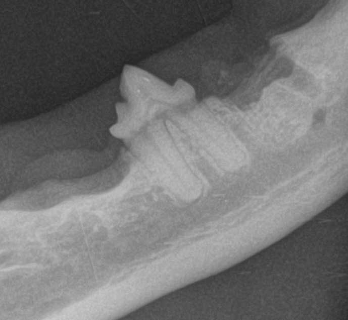
On anesthetized oral examination, tooth resorption is confirmed with the use of a dental explorer instrument and dental x-rays. X-rays are imperative for two reasons: 1- they reveal more lesions than oral examination alone and 2- they determine what the treatment will be. Lesions are classified as either type 1, 2 or 3 based on the progression of the disease. Type 1 lesions (often associated with periodontal inflammation) involve variable amounts of loss of the tooth structure, but the root and periodontal ligament remain intact. Type 2 lesions involve loss of normal root structure and the root structure takes on the radiographic appearance of alveolar bone. Multi-rooted teeth can display signs of both type 1 and 2 lesions and are therefore classified as type 3. Defining the type of lesion is how we determine how to treat resorption.
How do I prevent resorption from happening in my cat or dog?
Without knowing the cause of resorption, we cannot recommend any strategies for prevention other than daily brushing (which may or may not be affective). For this reason, if a patient has been diagnosed with resorption in the past, we recommend annual examinations, cleanings, and dental radiographs. Additionally, the fact that tooth resorption is so common in feline patients indicates that full-mouth dental radiographs should be part of every dental procedure in cats. As noted above, dental radiographs are vital in the diagnosis and treatment of tooth resorption.
Even though resorption is difficult to prevent, brushing is always a good idea to prevent periodontal disease and to keep an eye out for sore spots in your cat or dog’s mouth!
What is the treatment for tooth resorption?
Treatment of tooth resorption involves extraction of all affected dentition. We know that our domestic cats can function very normally with few to no teeth, so it is important that we not avoid treating this painful condition for fear of tooth loss. If we let the process continue, the tooth will eventually be lost regardless, it will just be a very long and painful process. In the past, restoration of these lesions with materials like glass ionomer has been attempted, but this is unsuccessful because the lesions progress regardless of the restoration.
The type of extraction will be determined by the type of lesion that was diagnosed on x-ray (as discussed above). Type 1 lesions require full extraction of all crown and root substance. Type 2 lesions involve removal of just the portion of crown and root that remain. This procedure is often called a crown amputation because of the loss of the root substance and subsequent inability to remove an entire intact root. The roots in type 3 lesions are treated individually by either extraction or crown amputation based on the presence or absence of root substance.